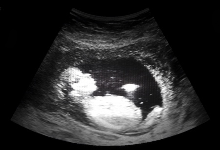
Ultrasound Exams
 You will obtain a few ultrasounds throughout the pregnancy.
- First trimester to establish or confirm the dating
- An anatomy ultrasound at 18 – 20 weeks
- You may receive additional ultrasounds if medically necessary
What does an ultrasound show?
An ultrasound creates pictures of the internal organs of the body from sound waves. This is no radiation involved. The sound waves are directed into a specific area of the body through a transducer.
The sound waves hit tissues, body fluids and bones. Waves then bounce back, like echoes, and are converted into pictures of the fetal parts.
The images appear on a computer screen. The dark areas indicate liquid like amniotic fluid. Gray or light areas show denser material like tissue. And white areas indicate bone.
The type of ultrasound that is used most often combines still pictures to show movement like the many single frames put together to make a movie. This is called real-time ultrasound.
When is ultrasound used in obstetrics?
Ultrasound is used in obstetrics to examine the growing fetus inside the woman’s uterus. A standard ultrasound exam can provide information about the fetus’ health and well-being including…
- Approximate fetal age
- Rate of fetal growth
- Placental location
- Fetal position, movement, breathing and heart rate
- Amount of amniotic fluid in the uterus
- Number of fetuses
- Some birth defects
